Anatomy Of Ribs And Muscles - Rib Wikipedia : It also serves as a connection point for other bones and muscles.
byAdmin-
0
Anatomy Of Ribs And Muscles - Rib Wikipedia : It also serves as a connection point for other bones and muscles.. The superior fibres originate from the spinous processes of the c7 to t3 vertebrae and attach to the superior borders of ribs two to four. Serratus posterio r consists of two muscles that assist respiration; Human anatomy drawing human figure drawing anatomy study anatomy art anatomy reference figure drawing reference pose reference anatomy bones body anatomy. The direction of the fibres parallels that of the innermost intercostal. The more anterior your cut, the more spinalis you'll find in the steak.
The eleventh and twelfth ribs have only one articular facet with no neck. Muscles also connect from one rib to the next. Lying exposed between the protective bones of the superiorly located ribs and the inferiorly located pelvic girdle, the muscles of this region play a critical role in protecting the. It supports and binds other tissues in the body. Each spinal segment includes two vertebrae separated by an intervertebral disc, the nerves that leave the spinal column at each vertebra, and the small facet joints that link each level of.
Learn Muscle Anatomy Serratus Posterior Superior And Inferior from www.visiblebody.com Others attach indirectly because they are attached to the cartilage of the rib above. The human rib cage is made up of 12 pairs of ribs, some of which attach to a bony process in the front of the chest called the sternum. Its function is to elevate the ribs. Human anatomy drawing human figure drawing anatomy study anatomy art anatomy reference figure drawing reference pose reference anatomy bones body anatomy. It also serves as a connection point for other bones and muscles. These muscles function in respiration by moving the ribs, thereby changing the volume of the thoracic cavity. The three layers of intercostal muscle are the external intercostal muscles, internal intercostal muscles, and the innermost intercostal muscles. The part of the muscle is thought to depress the ribs.
The three layers of intercostal muscle are the external intercostal muscles, internal intercostal muscles, and the innermost intercostal muscles.
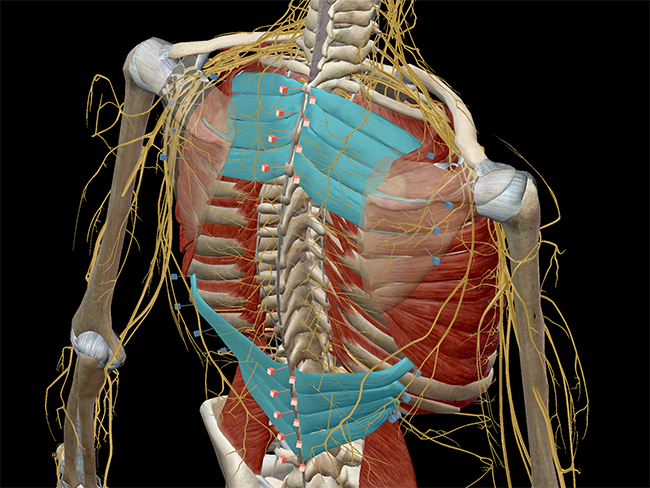
The subcostal muscles are found in the inferior portion of the thoracic wall. Serratus posterio r consists of two muscles that assist respiration; Muscles also connect from one rib to the next. The tenth rib has only one articular facet. Rib cage anatomy the rib cage shaped in a mild cone shape and more flexible than most bone sets is made up of varying elements such as the thoracic vertebra 12 human body ribs diagram the rib cage notasdecafe co. Human muscles · april 17, 2020. Rib below its origin, medial to the angle: Serratus posterior superior and serratus posterior inferior. The rectus abdominis is positioned between the ribs and the pubic bone at the front of the pelvis, and is actually made up of 8 distinct muscle bellies. A rib has a flat body, as you can see from the picture of the anatomy of the human rib cage. The first seven ribs attach directly to the sternum through cartilage that forms at the end of each rib. The human rib cage is made up of 12 pairs of ribs, some of which attach to a bony process in the front of the chest called the sternum. The slip arising from the sixth rib is the one most prominently seen on raising the arm away from the side, it passes.
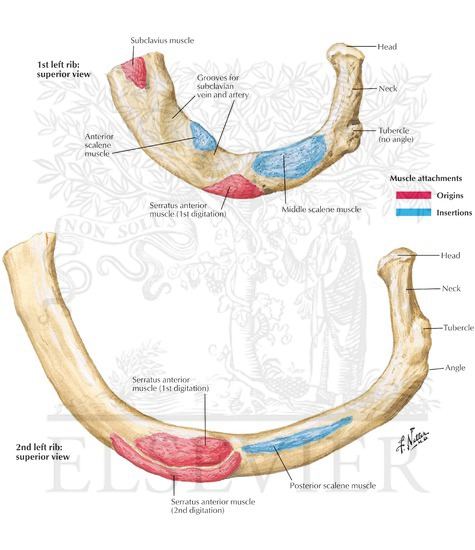
Serratus posterior superior and serratus posterior inferior. Details the different parts of the sternum (manubrium, sternal angle, xiphisternal joint) and the different muscle insertions (pectoralis major and sternocleidomastoid muscles). 202) passes from the side of the chest to the vertebral or posterior border of the scapula, arising by nine or ten digitations from the eight or nine upper ribs, the second having two. The upper edge is round and the lower sharp. The superior fibres originate from the spinous processes of the c7 to t3 vertebrae and attach to the superior borders of ribs two to four.
Muscle Attachments Of Ribs from www.netterimages.com These are fairly small and insignificant muscles: The rib section of beef spans from ribs six through twelve, and, obviously, hails from the rib section of the animal. Click the image to watch the anatomy of the rib cage video. The part of the muscle is thought to depress the ribs. They comprise of thin slips of muscle, which run from the internal surface of one rib, to second and third ribs below. Human anatomy drawing human figure drawing anatomy study anatomy art anatomy reference figure drawing reference pose reference anatomy bones body anatomy. These muscles function to move the shoulder girdle, spine, thorax, and pelvis and assist in respiration. A rib has a flat body, as you can see from the picture of the anatomy of the human rib cage.
These muscles function to move the shoulder girdle, spine, thorax, and pelvis and assist in respiration.
With the upper ribs, closer to the nodule (and in the case of lower ribs, a little further from the nodule) they are curved and have a rough surface that connects them with muscles, angulus costae. Deep cervical a., intercostal aa. The tenth rib has only one articular facet. It supports and binds other tissues in the body. They comprise of thin slips of muscle, which run from the internal surface of one rib, to second and third ribs below. Rib cage anatomy the rib cage shaped in a mild cone shape and more flexible than most bone sets is made up of varying elements such as the thoracic vertebra 12 human body ribs diagram the rib cage notasdecafe co. Click the image to watch the anatomy of the rib cage video. The second rib is thin, long, and has a tuberosity on its superior surface for the attachment of the serratus anterior muscle. Lying exposed between the protective bones of the superiorly located ribs and the inferiorly located pelvic girdle, the muscles of this region play a critical role in protecting the. Rib eye steaks are mainly composed of the longissimus dorsi muscle (the eye portion of the steak) and the spinalis dorsi muscle. Nov 23, 2020 · the internal intercostal muscles are the deeper set of muscles and depress the ribs to compress the thoracic cavity and force air to be exhaled from the lungs. The first rib is atypical because it is wide and short, has two costal grooves, and one articular facet. Details the different parts of the sternum (manubrium, sternal angle, xiphisternal joint) and the different muscle insertions (pectoralis major and sternocleidomastoid muscles).
It also serves as a connection point for other bones and muscles. Inserts into superior surface of first or second rib provide stability to the head and facilitate rotation; The slip arising from the sixth rib is the one most prominently seen on raising the arm away from the side, it passes. Lying exposed between the protective bones of the superiorly located ribs and the inferiorly located pelvic girdle, the muscles of this region play a critical role in protecting the. These are fairly small and insignificant muscles:
Introduction Anatomy Thoracic The Gap Physio from images.squarespace-cdn.com Related posts of muscle anatomy ribs muscle anatomy posterior. Serratus posterio r consists of two muscles that assist respiration; The tenth rib has only one articular facet. The rectus abdominis runs between the ribs and the pubic bone and supports movements between the rib cage and the pelvis. The direction of the fibres parallels that of the innermost intercostal. 202) passes from the side of the chest to the vertebral or posterior border of the scapula, arising by nine or ten digitations from the eight or nine upper ribs, the second having two. The slip arising from the sixth rib is the one most prominently seen on raising the arm away from the side, it passes. The second rib is thin, long, and has a tuberosity on its superior surface for the attachment of the serratus anterior muscle.
It also serves as a connection point for other bones and muscles.
Others attach indirectly because they are attached to the cartilage of the rib above. The eleventh and twelfth ribs have only one articular facet with no neck. The slip arising from the sixth rib is the one most prominently seen on raising the arm away from the side, it passes. Related posts of muscle anatomy ribs muscle anatomy posterior. Muscles also connect from one rib to the next. Über 7 millionen englischsprachige bücher. They comprise of thin slips of muscle, which run from the internal surface of one rib, to second and third ribs below. The three layers of intercostal muscle are the external intercostal muscles, internal intercostal muscles, and the innermost intercostal muscles. The rectus abdominis is positioned between the ribs and the pubic bone at the front of the pelvis, and is actually made up of 8 distinct muscle bellies. Human anatomy drawing human figure drawing anatomy study anatomy art anatomy reference figure drawing reference pose reference anatomy bones body anatomy. Serratus posterio r consists of two muscles that assist respiration; At the chest, many rib bones connect to the sternum via costal cartilage,. Lying exposed between the protective bones of the superiorly located ribs and the inferiorly located pelvic girdle, the muscles of this region play a critical role in protecting the.
The muscles of the abdomen, lower back, and pelvis are separated from those of the chest by the muscular wall of the diaphragm, the critical breathing muscle anatomy of ribs. Lying exposed between the protective bones of the superiorly located ribs and the inferiorly located pelvic girdle, the muscles of this region play a critical role in protecting the.